3 Effective Tools for Oncology Drug Development
This article Is brought to you by Noble Life Sciences, a leading preclinical contract research organization (CRO) specializing In oncology drug development.
| Noble Life Sciences is an Industry Partner who values community just as much as we do. They are a contract research organization (CRO) that provides a full continuum of preclinical drug, vaccine and medical device development services, from initial product discovery to GLP-compliant studies. Enjoy the read and be sure to get in touch if you have an idea or perspective you’d like to share. BioBuzz welcomes guest posts and contributing writers with expertise on topics that are of interest. |
In the realm of oncology drug development, both biopharma companies and their trusted partners, contract research organizations (CROs), wield an extensive arsenal of tools. These tools guide drugs from their initial identification stage to full commercialization. Noble Life Sciences, among other preclinical oncology CROs, employs many of these tools on a daily basis. In this article, we delve into three effective tools that have become integral to the oncology drug development process. In this article, Noble Life Sciences shares how they use them with you and offers its expertise and firsthand insights, contributing to the progress of biotech, biopharma, and pharma companies in advancing their preclinical programs.
1. Cell Viability Assays for Drug Screening:
One of the cornerstone tools in oncology drug development is the cell viability assay. The MTT assay, which assesses the metabolic activity of cells and works on a straightforward principle – only viable cells exhibit active metabolism, converting MTT (3-(4,5-dimethylthiazol-2-yl)-2,5-diphenyl-2H-tetrazolium bromide) into formazan, which appears as a purple compound in colorimetric tests. By measuring the amount of formazan produced using a spectrophotometer, scientists can determine the number of viable cells in a culture.
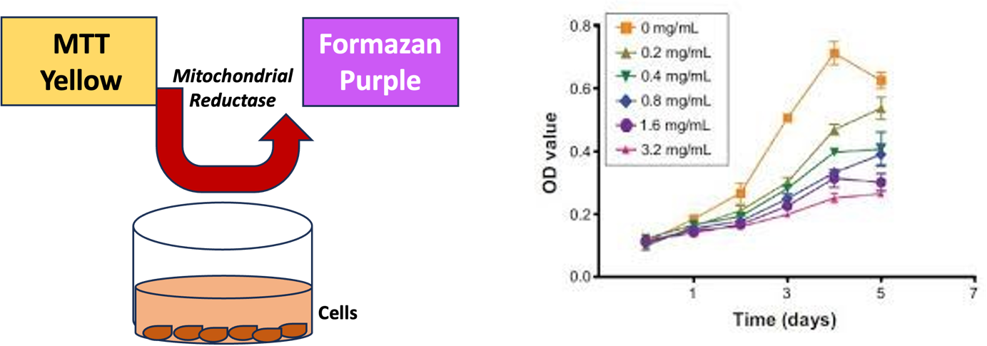 |
| Figure 1. MTT assay: MTT (Yellow) when added to the media containing cells, gets taken in, converted into purple formazan by metabolic process and released into the medium by the cells. This can be quantified in spectrophotometer at 490 nm absorbance for the cell proliferation and/ or metabolic activity of the cells. A useful assay to study cell proliferation in test compound screening, A sample image showing the cell proliferation data for MTT assay, where cell proliferation without and with different concentrations of a drug were measured by MTT assay (Lin Q et al., Int. J of Oncology, 53: 246-256). |
The MTT assay has utility in measuring cell proliferation and viability, drug cytotoxicity, and mitochondrial and metabolic cellular activity. For instance, in oncology drug development, it helps assess the potency of a new version of an oncology drug compared to the original. It also aids in predicting a drug’s efficacy against malignancies.
Florento et al. have noted that “The test is easy to perform in hematological malignancies and is adaptable for high throughput of samples…. This class of assay is highly accurate for predicting drug resistance, where its predictive value for drug sensitivity depends on the type of disease and drug or drug combination used.” The MTT assay, along with similar tests, assists researchers in selecting chemotherapeutic drugs with the highest clinical effectiveness while excluding ineffective candidates.
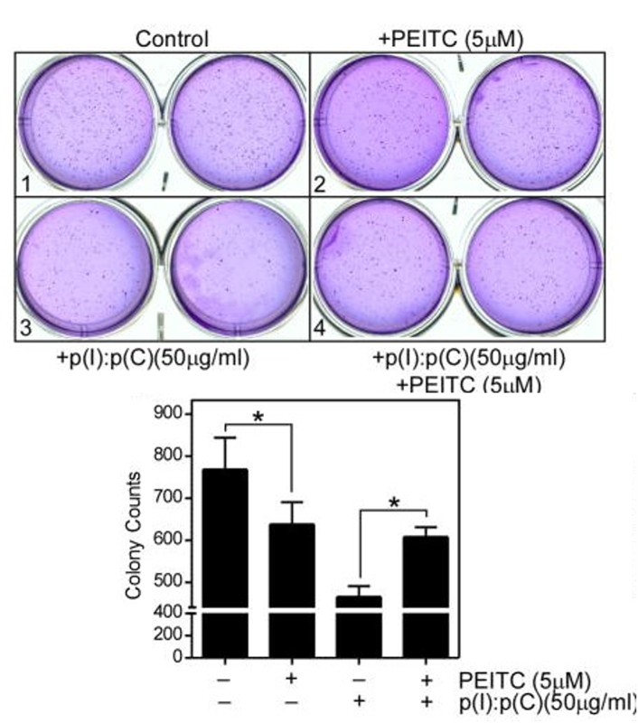
2. Anchorage-Independent Growth Assays:
Anchorage-independent growth, a characteristic often associated with cancer cells, refers to the ability of cells to grow without being tethered to a solid surface. To evaluate this capability in vitro, scientists use the soft agar colony formation assay, a well-established method that is more cost-effective and easier to perform than in vivo tests involving laboratory animals. Furthermore, some malignant cells do not form tumors when transplanted into animals.
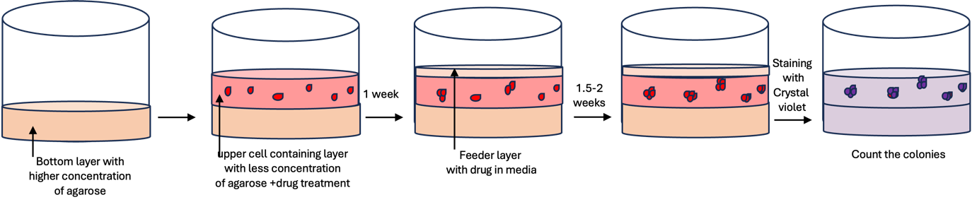 |
In this assay, cells are dispersed onto a culture plate and grown either with “feeder” cells or a conditioned medium containing appropriate growth factors. It provides data specifically on colony formation. Normal cells, incapable of anchorage-independent growth due to anoikis (a form of apoptotic death), serve as a reference. Variations of this assay, such as using specialized agar to collect viable cells for protein or DNA sampling, have been developed for various purposes.
 |
For instance, in the study of medulloblastoma, a common malignant childhood brain cancer, a 3D 384-well agar colony formation assay was created to assess the growth of medulloblastoma cells. This assay used two fluorescent substrates, resazurin and glycyl-phenylalanyl-aminofluorocoumarin (GF-AFC), to measure cell viability. It allowed researchers to screen for compounds affecting medulloblastoma cell growth in a matrix similar to three-dimensional tumor growth in vivo.
3. Cell Migration and Invasion Assays:
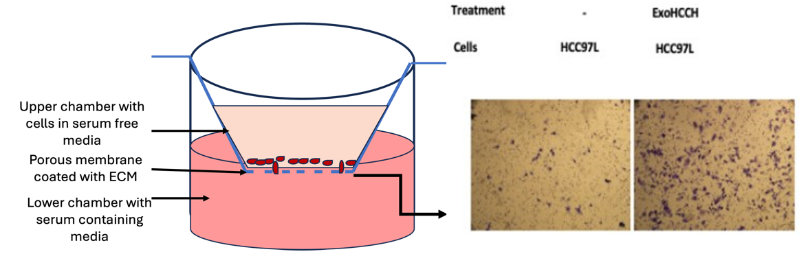
The ability of cancer cells to migrate and invade other tissues is critical in understanding cancer progression and recurrence. Two primary assays for evaluating this dissemination are the Boyden chamber assay and the scratch wound assay, each with its own strengths and limitations.
- Boyden Chamber Assay: This assay employs a chamber with two separate compartments, separated by a microporous membrane, filled with medium. Cells are placed in the upper compartment and allowed to migrate through the membrane into the lower compartment, which contains chemotactic agents. After fixing and staining the membrane, the number of cells that migrated to the lower side is counted. In cancer research, it can evaluate the invasiveness of tumor cells by coating the membrane with various extracellular matrix proteins.
 |
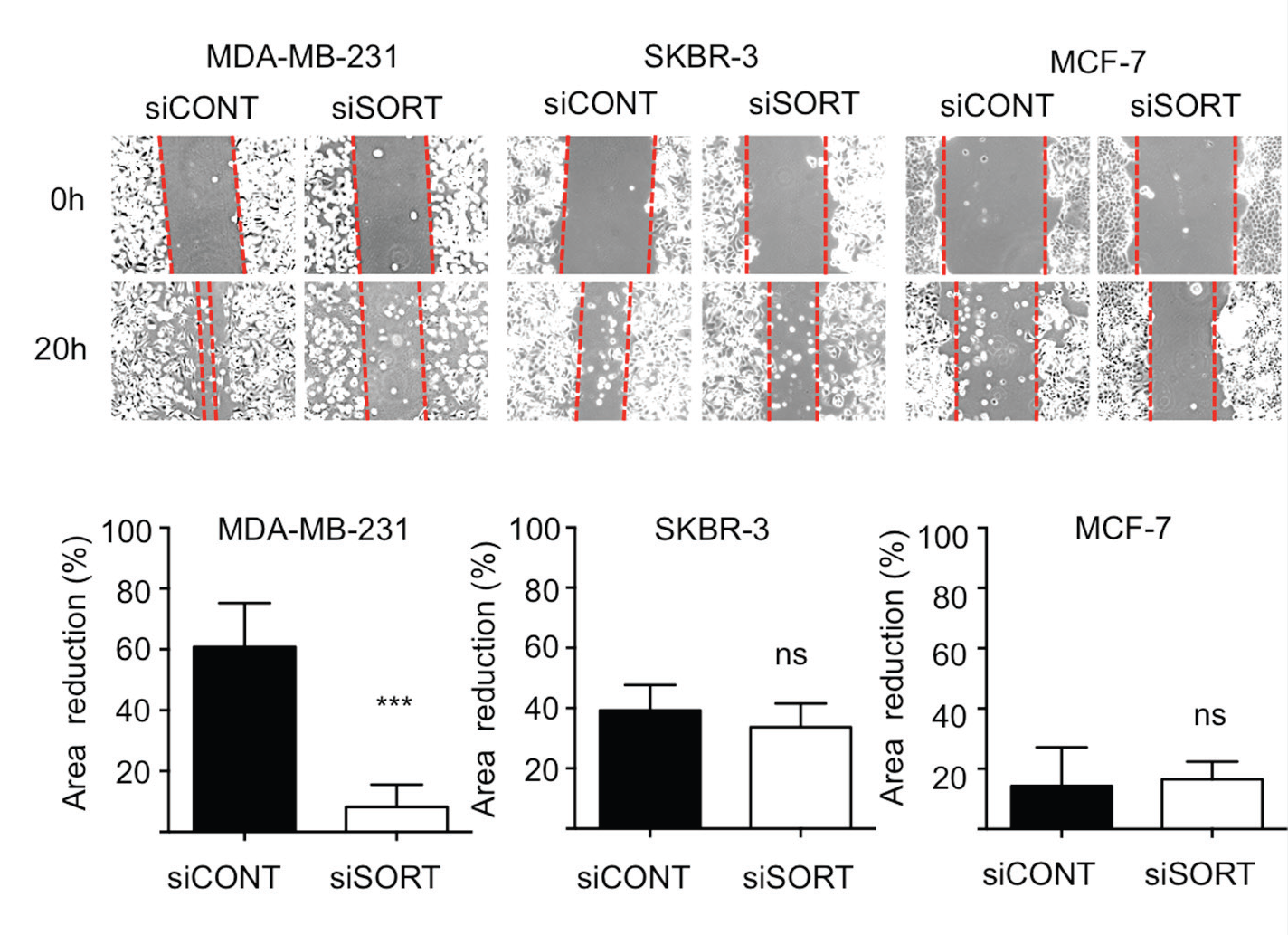
- In Vitro Scratch Wound Assay: This method measures cell migration by creating a “wound” in a monolayer of cells using a commercial wounding device. Images are capture for cell migration from both sides of the wound. Although the scratch wound assay traditionally had low reproducibility, newer approaches, particularly with microscopy, have improved its reliability.
 |
Experienced CROs, like Noble Life Sciences, boast a diverse toolbox that aids pharma companies in designing and developing oncology therapeutics. These three tools represent just a fraction of the vast array at their disposal, with others including humanized rodent models for immune-oncology studies, oncolytic virus-mediated cancer therapy models, patient-derived xenografts, cell line-derived xenografts, and many more.
Noble Life Sciences can screen a diverse collection of cells scanning multiple indications focused on client needs. The results from this panel can be correlated with the cellular and molecular characteristics of these cancer cell lines, facilitating the identification of molecular biomarkers that align with the activity of the biopharma company’s drug candidates.
With this diverse toolkit and advanced techniques, CROs play a pivotal role in helping biopharma companies navigate the drug development journey efficiently, providing the essential data needed for confident decision-making.
Sources
1. Ghasemi M, Turnbull T, Sebastian S, Kempson I. The MTT Assay: Utility, Limitations, Pitfalls, and Interpretation in Bulk and Single-Cell Analysis. Int J Mol Sci. 2021;22:12827. https://www.ncbi.nlm.nih.gov/pmc/articles/PMC8657538/
2. Florento L, Matias R, Tuaño E, Santiago K, dela Cruz F, Tuazon A. Comparison of Cytotoxic Activity of Anticancer Drugs against Various Human Tumor Cell Lines Using In Vitro Cell-Based Approach. Int J Biomed Sci. 2012;8(1):76-80. https://www.ncbi.nlm.nih.gov/pmc/articles/PMC3614850/
3. Afify SM, Seno M. Anchorage-independent Cell Growth Assay for Cancer Stem Cells: Tumirogenic Assay in Vitro. In: Methods in Cancer Stem Cell Biology. Springer, Singapore. 2023. https://link.springer.com/chapter/10.1007/978-981-99-1331-2_16#
4. Borowicz S et al. The Soft Agar Colony Formation Assay. J Vis Exp. 2014;(92):e51998. https://www.jove.com/t/51998/the-soft-agar-colony-formation-assay
5. Sedeeq M, Maklad A, Gueven N, Azimi I. Development of a High-throughput Agar Colony Formation Assay to Identify Drug Candidates against Medulloblastoma. Pharmaceuticals (Basel). 2020;13(11):368. https://www.ncbi.nlm.nih.gov/pmc/articles/PMC7694510/
6. Guy J-B, et al. Evaluation of the Cell Invasion and Migration Process: A Comparison of the Video Microscope-based Scratch Wound Assay and the Boyden Chamber Assay. J Vis Exp. 2017;129:e56337. https://pubmed.ncbi.nlm.nih.gov/29286429/
###